On Hadatomo™ Z, high-resolution ultrasonic sensor moves and scans measurement area, obtaining photoacoustic signals and ultrasonic signals. In each measurement point, two-wavelengths photoacoustic imaging and ultrasound imaging are conducted in high speed, then moves to the next point. After measurement is finished, measurement data of two kinds of photoacoustic images and ultrasound images can be saved. The measured data can be displayed as 3D images.
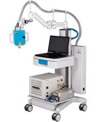
Hadatomo™ Z Photoacoustic Microscope WEL5200
Noninvasive, High-Speed 3D Imaging of Blood Vessels in the Dermis
The Hadatomo™ Ζ simultaneously images the oxygen saturation of blood vessels with two wavelengths of optical ultrasound, and images the skin texture, pores, and skin structures such as sebaceous glands with ultrasound.
- Features/Specifications
- Measurement Example
- Measurement Principle
- Research Achievement
- Download Leaflet
- Contact
Feature
Multi-modality Imaging
Ultrasound that visualizes the skin structure based on the differences in tissue hardness, and optical ultrasound, which selectively images melanin and blood vessels based on their absorption characteristics, are integrated into one system.
Quasi-real-time imaging
The photoacoustic wave images and the ultrasonic wave images are measured simultaneously, with a minimum measurement time of 40 seconds (Range: 6 mm x 6 mm x 3 mm (depth)) and maximum time of 210 seconds (Range: 9 mm x 9 mm x 3 mm (depth)).
High-resolution images obtained with easy operation
An ultrasonic sensor developed especially for this system provides a high-resolution crosssection view. Focus points can be easily located by observing the cross-section.
Flexible design with easy operation and portability
The compactly designed system, equipped with a measurement unit mounted on a flexible arm, is installed on a system unit with caster wheels, easy to set up to measure various parts of the body.
Label-free
Applying a small amount of water (acoustic coupling agent) to the measurement area is enough for measurement.
Non-invasive measurement is possible without any contrast agent.
Acquires data for 3-dimensional imaging
Measurement data are displayed as 2-dimensional cross-section images in real-time, and acquisition of 3-dimensional data images is possible. 3-dimensional image analysis is possible with customer-provided rendering software.
Specification
System
| Measurement | 2D(Tomography), 3D |
|---|---|
| Sampling frequency | 500 MHz |
| Dimensions | Approx. 610 (W) × 730 (D) × 1,400 (H) |
| Weight | < 135 kg |
3D image
| Measurement area(x, y) | (1) 6 x 6 mm (2) 9 x 9 mm |
|---|---|
| Measurement depth(z) | 3 mm (designated signal acquisition range) |
| Scanning step | 15 μm / 30 μm |
| Measurement time(15 μm) | (1) 210 s (2) 420 s |
| Measurement time(30 μm) | (1) 70 s (2) 140 s |
2D image
| Measurement area(x, ) | 6 mm |
|---|---|
| Measurement depth(z) | 3 mm |
| Scanning step | 15 μm / 30 μm |
| Flame rate | 1 fps |
Photoacoustic imaging
| Wavelength | (A) 532/556 nm (B) 575/650 nm *1 |
|---|---|
| Horizontal image display | 15 μm |
| Axial image display | 12 μm *2 |
| Pulse energy |
(A)Below 16 µJ/pulse
(B)Below 18 µJ/pulse (575 nm)
Below 14 µJ/pulse (650 nm) *3 |
| Pulse width | Below 10 ns |
| Repetition rate | 1000 Hz *4 |
Ultrasound imaging
| Horizontal image display | 15 μm |
|---|---|
| Axial image display | 12 μm *5 |
| Transmission repetition rate | 1000 Hz |
Ultrasound sensor
| Center frequency | 60 MHz ± 20 % |
|---|
*1: Select one of the following sets: 532/556 nm, or 575/650 nm
*2: Depending on ultrasound velocity
*3: Averaged value
*4: 1000 Hz per 1 wavelength
*5: Depending on ultrasound velocity
* Specifications may change without notification.
* This product is categorized as a Scientific Instrument. It is not approved for use in diagnosis of diseases.
| 3D | 2D | |
|---|---|---|
Face
 |
 575 / 650 nm Laser Unit  |
 575 / 650 nm Laser Unit  |
Upperarm
 |
 575 / 650 nm Laser Unit  |
 575 / 650 nm Laser Unit  |
Hair
 |
575 / 650 nm Laser Unit  |
575 / 650 nm Laser Unit  |
| Focused on Eponychium | Focused on Nail Bed | |
|---|---|---|
 |
 Vascular Network of Eponychium |
 Vascular Network of Nail Bed |
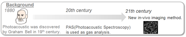
The photoacoustic effect was discovered by Graham Bell in the 19th century.
In the 20th century, its applications included gas analysis instruments. With the technical innovation on laser, ultrasonic wave and PC, its applications for in-vivo imaging is being developed in the 21st century.
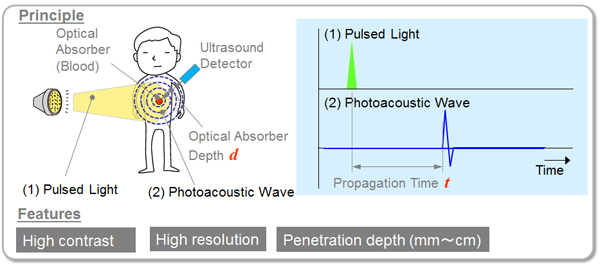
When pulses of light with a certain wavelength are irradiated into tissue, material with absorption characteristics to the wavelength (hemoglobin) selectively absorbs their energy and causes adiabatic expansion.
It generates thermoelastic waves, propagating within the tissue as ultrasonic waves, so they can be captured as signals, by sensors installed on the surface of the body.
The propagation time of the ultrasonic waves give accurate depth information, and they can visualize images of material distribution. Also, by using the absorbing characteristics of light, high contrast images can be obtained.
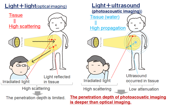
Tissue has high-scattering characteristics against light. Therefore, the optical imaging has limitations on penetration depth, because the light radiated to the tissue, and the light reflected in the tissue, are both affected by the scattering characteristics.
In contrast, on the ultrasound imaging, the irradiated light for the tissue are affected with the scattering characteristics, but the generated ultrasonic waves keep high-propagation characteristics within the tissue, achieving better penetration depth compared to the optical imaging.
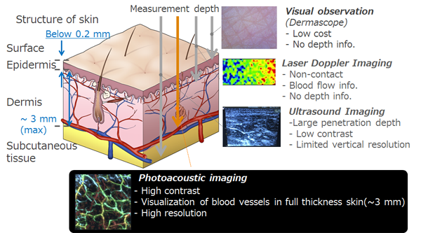
Ultrasonography visualizes structure of tissue noninvasively, utilizing the hardness difference of the living tissues.
However, it is difficult to visualize blood vessels within the dermis, because hardness differences are hardly found in shallow area of tissues.
In contrast, optical imaging, such as microscopy can obtain high-resolution images, but their immersion depth is less than 1 mm. This means in the shallow area of tissues, in the range of mm to cm, there are areas where existing technology could not visualize.
Photoacoustic imaging is a novel imaging method to visualize the area of mm to cm.
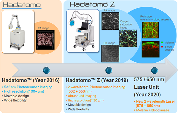
Advantest has been researching and developing the photoacoustic imaging technique. In 2016, we launched Photoacoustic microscope Hadatomo™ and in 2019, we launched high-resolution version of Photoacoustic microscope, Hadatomo™ Z.
Hadatomo™ Z can use two-wavelength photoacoustic imaging and ultrasound imaging. Using the two-wavelengths (532/556 nm) photoacoustic imaging, calculation such as oxygen saturation becomes possible, and superimposing the ultrasound images and the photoacoustic images can clearly display blood vessels running within the dermis. In 2020, we developed new 2-wavelengths lasers (575/650 nm) for Hadatomo™ Z. The image obtained with the 650 nm wavelength can visualize melanin, while the image obtained with the 575 nm wavelength can visualize vascular network, so the melanin and the vascular network can be displayed separately.
The system is a valuable new research tool for dermatology researchers.
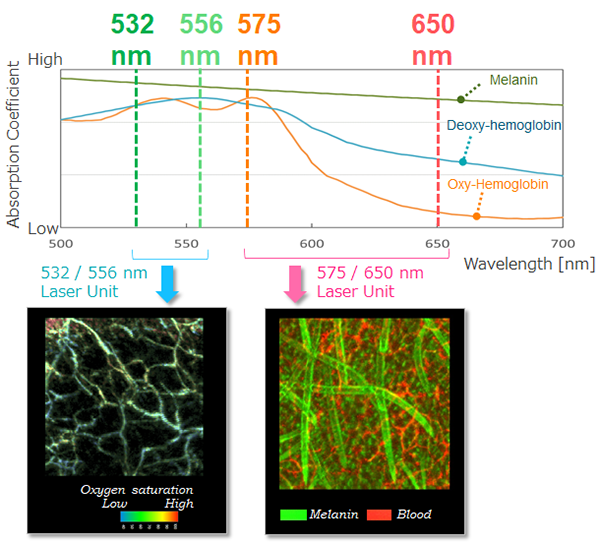
Absorption coefficient of the oxy-hemoglobin and the deoxy-hemoglobin are different in each wavelength. By utilizing the 532 + 556 nm Laser Unit, we can calculate the oxygen saturations from the measured photoacoustic signals. In contrast, absorption coefficient of melanin is as high as hemoglobin, so it is difficult to distinguish the melanin and the blood vessels.
In the wavelength of 650 nm, absorption coefficient of the hemoglobin is lower, so that selective visualization of melanin is possible. In the image obtained with the 575 nm wavelength, both melanin and blood vessels are similarly visualized. However, by calculating the difference between the signals obtained with the 650 nm and 575 nm wavelengths, blood vessels can be extracted. By superimposing both images, 3D images of the melanin and the blood vessels can be created.
Using the two kinds of photoacoustic images, oxygen saturation can be measured, and by superimposing the photoacoustic images and ultrasound images, the blood vessels inside the dermis can be observed. Also, the raw data can be used for numerical analysis.
Hadatomo™ Z has castor wheels, to be relocated easily. Various body regions can be measured by its flexible movable arm.
- Taiichiro Ida, Hideaki Iwazaki, Toshiyuki Omuro, Yasushi Kawaguchi, Yasuyuki Tsunoi, Satoko Kawauchi, and Shunichi Sato, “Lensless high-resolution photoacoustic imaging scanner for in vivo skin imaging,” Optical Review 25(1) DOI:10.1007/s10043-017-0384-1 (2018).
- 伊田 泰一郎,川口 康,大倉 津矢子,川内 聡子,津田 均,斎藤 大蔵,佐藤 俊一,岩井 俊昭, “リアルタイム光音響イメージング法による移植皮膚の生着モニタリング:ラット自家皮膚・同種皮膚移植モデルによる実験,” 熱傷 42(2), pp. 57-63 (2016).
- Taiichiro Ida, Hideaki Iwazaki, Yasushi Kawaguchi, Satoko Kawauchi, Tsuyako Ohkura, Keiichi Iwaya, Hitoshi Tsuda, Daizoh Saitoh, Shunichi Sato, and Toshiaki Iwai, “Burn depth assessments by photoacoustic imaging and laser Doppler imaging,” Wound Repair and Regeneration DOI: 10.1111/wrr.12374 (2015).
- Taiichiro Ida, Yasushi Kawaguchi, Satoko Kawauchi, Keiichi Iwaya,Hitoshi Tsuda, Daizoh Saitoh, Shunichi Sato, and Toshiaki Iwai, “Real-time photoacoustic imaging system for burn diagnosis,” Journal of Biomedical Optics 19(8):086013 (2014).
- Yasushi Kawaguchi, Hideaki Iwazaki, Taiichiro Ida, Taiji Nishi, Yukari Tanikawa, and Naotaka Nitta, “New polymer-based phantom for photoacoustic imaging,” Proc. SPIE 8945, Design and Performance Validation of Phantoms Used in Conjunction with Optical Measurement of Tissue VI, 89450A (2014).
- Taiichiro Ida, Yasushi Kawaguchi, Satoko Kawauchi, Keiichi Iwaya, Daizoh Saitoh, Shunichi Sato, and Toshiaki Iwai, “Real-time photoacoustic imaging system for clinical burn diagnosis,” Proc. SPIE 8581, Photons Plus Ultrasound: Imaging and Sensing, 858109 (2013).
- 川口康,伊田泰一郎,川内聡子,齋藤大蔵,佐藤俊一,“光超音波技術が拓く新たな世界,” BIO Clinica 30(2), pp. 69-75 (2015).
- 伊田泰一郎,川口康,川内聡子,岩屋啓一,齋藤大蔵,佐藤俊一,岩井俊昭,“リアルタイム光超音波イメージング・システムによる熱傷深度診断,” ADVANTEST Probo No.41, pp. 24-29 (2014).
- 川口康,伊田泰一郎,保坂智也,岩崎秀明,“光超音波イメージング・システム,” 超音波テクノ, 25(4), pp. 98-102 (2013).
- 川口康,伊田泰一郎,川内聡子,齋藤大蔵,佐藤俊一, “光超音波イメージング:新たなモダリティ開拓への挑戦~光音響技術を誰でも使えるものに~,” 日本レーザー医学会誌 33(4), pp. 374-379 (2013).
- 伊田泰一郎, “光超音波イメージング” OPTO NEWS 8(1), pp. 12-16 (2013).
| Model | Download | |
|---|---|---|
| WEL5200 | Photoacoustic Microscope | Leaflet (English) |
| WEL5200 | Photoacoustic Microscope | Leaflet (Chinese) |
If you need further assistance, please use contact form clicking the link below.