
Key Features of New Technology
- Imaging of oxygen saturation distribution in the vascular network of skin
- Control of ultrasonic wave transmission and reception with dual-wavelength light source enables near-real-time in vivo imaging
TOKYO, Japan – October 12, 2018 – Leading semiconductor test equipment supplier Advantest Corporation (TSE: 6857) announced that as part of the “Innovative Visualization Technology to Lead to Creation of a New Growth Industry” project operated by Takayuki Yagi under the auspices of the Impulsing Paradigm Change through Disruptive Technologies Program (ImPACT), a program of Japan’s Council for Science, Technology and Innovation, an R&D group led by Professor Yoshifumi Saijo of Tohoku University and Noriyuki Masuda of Advantest has succeeded in developing in vivo skin imaging technology (1) that can simultaneously generate dual-wavelength photoacoustic images and ultrasound images.
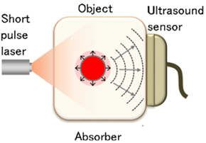
Photoacoustic imaging (2) is a method of imaging the interior of a living body by irradiating light into the body and measuring ultrasonic waves generated from blood or tissues that selectively absorb light energy. It is attracting interest as a new noninvasive imaging method suitable for measuring small blood vessels in the skin, which is difficult with conventional imaging techniques.
However, when using only photoacoustic imaging, even if microvessels in the skin measuring several tens of microns or less in diameter are imaged, it is impossible to ascertain which region of each layer in the skin they are in. In addition, it is possible to photoacoustically measure the oxygen saturation level of blood vessels (3) by using light sources of multiple wavelengths, but the movement of living bodies affects measurement results, so the use of this method has hitherto been limited to research applications such as animal experiments.
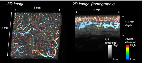
The newly developed in vivo imaging technology utilizes a focused ultrasonic sensor that can detect multiple ultrasonic signals. Thus, photoacoustic waves and ultrasonic waves can be measured with the same sensor, while signals are generated on two alternating wavelengths, allowing the detection of ultrasonic waves that image the microvascular network in the dermis as well as blood oxygen saturation (Fig. 1). A 6 mm square area of 2 mm depth can be imaged in about 4 minutes. Also, using the acquired data, mapping of oxygen saturation and the superposition of photoacoustic images and ultrasound images is possible.
Biopsy studies have proved that signs of skin aging such as spots and wrinkles are related to microvessels in the skin. The newly developed photoacoustic imaging method is expected to be used for monitoring of photoaging of the skin as well as other applications.
Blue indicates lower oxygen saturation of blood vessels, red higher.
These results were achieved with support from the following program:
Impulsing Paradigm Change through Disruptive Technologies Program (ImPACT)
- Program Manager :
- Takayuki Yagi
- R&D Program :
- Innovative Visualization Technology to Lead to Creation of a New Growth Industry
- R&D Topics :
- Development of ultra-high resolution photoacoustic imaging technology; development of micro-visualization system prototype
- R&D Director :
- Professor Yoshifumi Saijo, Graduate School of Biomedical Engineering, Tohoku University
- R&D Period :
- October 2014 – March 2019
- R&D Topics :
- Development of a micro-visualization system prototype
- R&D Director :
- Noriyuki Masuda, VP of New Concept Product Initiative, Advantest Corporation
- R&D Period :
- September 2016 – March 2019
This R&D team is working on the development of a micro-visualization system to achieve high-resolution imaging of the skin.
Comment from Program Manager Takayuki Yagi
In this program, we are working on a non-invasive method to achieve real-time 3D imaging of the vascular network and blood condition (oxygen saturation) of the living body, using a photoacoustic method to detect ultrasonic waves generated by laser irradiation. This method may be used for early diagnosis and monitoring of physical functions related to beauty and health. In addition, we are promoting the development of a non-destructive method to visualize deterioration and cracks in industrial materials.
Hitherto, photoacoustic imaging for measuring microvessels of several tens of micrometers or less and the blood condition of the skin has been limited to research applications such as animal experiments. Also, it was not possible to ascertain where the measured blood vessels were located inside the skin. Tohoku University and Advantest Corporation have succeeded in developing a superior photoacoustic 3D imaging technology by developing a more compact ultrasonic sensor than can simultaneously measure dual-wavelength photoacoustic images and ultrasonic images. This makes it possible to noninvasively image blood vessels and melanin in facial skin, opening up applications in the beauty field.

Background
In this program, researchers sought to develop new bio-imaging technology using photoacoustic technology. The team’s goal was to develop a micro-visualization system that achieves a microscopic lateral resolution of 30 μm or less, with the aim of imaging the vascular network in the dermis at high resolution.
Photoacoustic imaging can selectively image substances (such as blood and melanin) that have optical absorption properties, but it cannot distinguish between melanin and blood vessels, arteries and veins, etc. with a single wavelength light source, so information obtained is limited. Also, since the structure of the skin cannot be imaged, the surface of the skin and the thickness of the skin are “invisible,” and the location of blood vessels and melanin cannot be identified with photoacoustic images alone.
Photoacoustic imaging at multiple wavelengths and superposition of ultrasonic images can overcome these problems, but in order to utilize these methods for in vivo imaging, a high-speed system is required.
Research
To solve the problems described above, Tohoku University and Advantest Corporation set out to develop a micro-visualization system capable of acquiring dual-wavelength photoacoustic images and simultaneously acquiring ultrasound images.
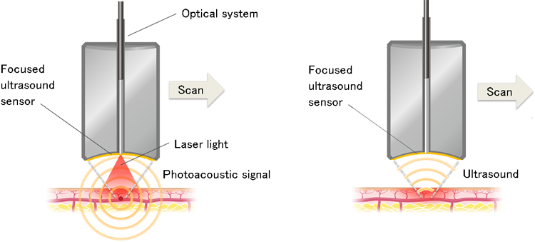
As a high-resolution photoacoustic imaging method, the team adopted a focused ultrasonic sensor for scanning. With this sensor, ultrasonic waves bounced off blood vessels, etc. can be collected on the surface of the sensor to obtain photoacoustic images (Fig. 2 (left)), and it is possible to acquire ultrasound images from the same sensor by collecting the ultrasound waves that were transmitted and reflected from it (Fig. 2 (right)). Moreover, by integrating an optical system with a laser light source and an ultrasonic sensor, the team was able to make the sensor much more compact, making it possible to bring it closer to facial skin.
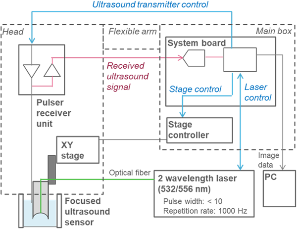
This scanning methods have the drawback that when scanning is performed separately for each wavelength, inevitable movements of the living body affect measurement results, and therefore it is necessary to scan with a sensor after performing dual-wavelength optical ultrasonic imaging and ultrasonic imaging at the measurement location. To accelerate this process, it is necessary to synchronize the dual-wavelength light source, ultrasonic transmission / reception, and sensor scanning in a single system. The system newly developed by the Tohoku University and Advantest researchers (Fig. 3) incorporates a dedicated board to control a dedicated-wavelength light source, ultrasonic sensor and XY stage. This has drastically shortened measurement times so that now a 6 mm square and 2 mm depth can be scanned in 15 μm steps for a total measurement time of about 4 minutes.
The system’s dual-wavelength light source can alternately irradiate pulsed light of 532 nm and 556 nm, and oxygen saturation data can be obtained from the difference between the obtained photoacoustic signals. By superimposing ultrasonic images on photoacoustic images, it is also possible to investigate correlation with information about blood vessel distance from the skin’s surface, pores, sebaceous glands, and so on.
Maximum permissible exposure (4) is stipulated as a laser safety standard for living bodies (eyes or skin). This system satisfies the MPE for the skin, so that in vivo imaging of facial skin etc. is possible. Thus, it has potential as a new tool for dermatological research and development, and as a new diagnostic device, as it can image the oxygen saturation of the vascular network in the dermis, which was impossible in the past, in combination with photoacoustic images.
In the future, the team will continue its research on the effectiveness of oxygen saturation measurement and work on practical applications for the system.
Notes
-
(1)In vivo imaging: Imaging of a living body
-
(2)Photoacoustic imaging: A method of detecting and imaging the ultrasonic waves generated by thermal expansion of an absorber that absorbs light (a phenomenon known as the photoacoustic effect) by irradiating the absorber with a pulsed laser beam. (See figure at right.)
-
(3)Oxygen saturation: The proportion (%) of oxygenated hemoglobin of total hemoglobin in the blood. In photoacoustic imaging, we can calculate oxygenated hemoglobin concentration and de-oxygenated hemoglobin concentration from acoustic pressures obtained at two wavelengths to calculate oxygen saturation.
-
(4)Maximum permissible exposure (MPE): Calculated as 10% of the dose that has a 50% chance of creating damage under worst-case conditions.
Enquiries
<R&D>
Professor Yoshifumi Saijo
Graduate School of Biomedical Engineering, Tohoku University
Tel:022-795-7148 Fax:022-795-7149
Advantest Corporation PR Section
Tel:03-3214-7500
<ImPACT>
ImPACT Program Promotion Office, Cabinet Office
Tel:03-6257-1339
<ImPACT Programs & Program Managers>
ImPACT Program Promotion Office
Tel:03-6380-9012 Fax:03-6380-8263
<Press Coverage>
Graduate School of Biomedical Engineering, Tohoku University
Tel:022-795-7491
Advantest Corporation PR Section
Tel:03-3214-7500
Japan Science & Technology Agency PR Section
Tel:03-5214-8404 Fax:03-5214-8432
-
Note:All information supplied in this release is correct at the time of publication, but may be subject to change.